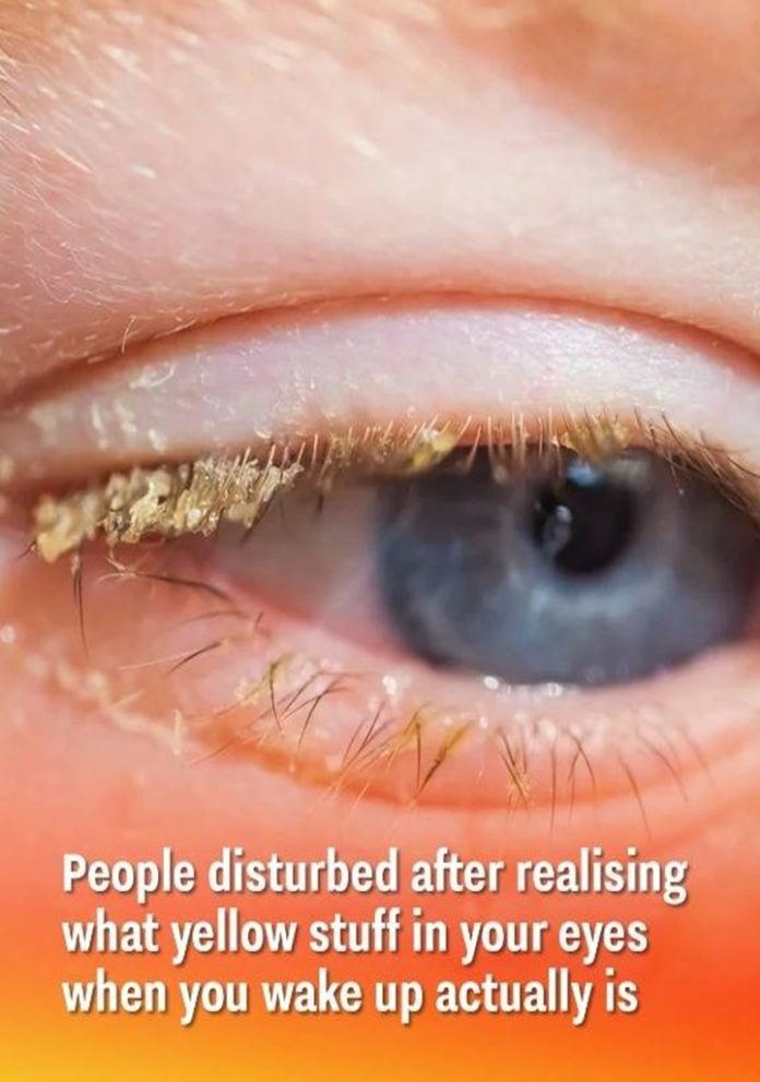
The daily act of waking up often begins with a simple, universally shared ritual: the gentle clearing of the eyes. We reflexively wipe away the yellowish, sometimes hardened, crusty bits that invariably gather in the inner corners of our eyes while we sleep. This common residue, known colloquially as “sleep,” “eye boogers,” or “eye gunk,” is so familiar and seemingly trivial that it is barely registered. Yet, despite how normal and mundane this phenomenon is, few truly understand what this discharge actually contains—or, more significantly, its vital importance to maintaining the continuous health and defense of the ocular system. A recent surge of public interest, fueled by neuroscientists like Andrew Huberman, has highlighted the complexity of this everyday process, revealing that what many view as a minor, slightly “gross” nuisance is actually a clear, powerful sign of the eye’s natural, sophisticated defense system working tirelessly and successfully overnight.
I. Unmasking the Composition: The Ingredients of Morning Eye Crust
The dried residue found in the eyes upon waking is far more complex than a simple accumulation of external dust or dried tears. It is a carefully orchestrated biological compound—a byproduct of the eye’s essential maintenance and immune functions. Dr. Rachel Clemmons, a leading ophthalmologist, clarifies: “Tears contain complex antimicrobial proteins, and mucus acts as a primary barrier. The discharge we see is a visible sign that the eye’s immune system has successfully neutralized bacteria and collected debris that might have caused infection.”
The crust is a functional composite of the eye’s primary defense components, each playing a vital role in protection:
1. Mucus (Mucin)
- Source: Produced by specialized goblet cells located within the conjunctiva (the clear membrane covering the white of your eye and inner eyelid).
- Function: Mucus, or mucin, is a sticky, gelatinous substance designed to trap minute particles, airborne debris, and environmental irritants, preventing them from lodging on the delicate corneal surface. It is the initial, protective glue of the tear film.
2. Oils (Lipids)
- Source: Secreted by the meibomian glands, which line the edges of the eyelids.
- Function: These oils (lipids) form the critical outer layer of the tear film. Their hydrophobic nature is essential for preventing the watery tear layer beneath from evaporating too rapidly. This ensures sustained moisture and provides smooth, friction-free lubrication every time the eye moves or the eyelid blinks.
3. Tears (Aqueous Fluid)
- Source: Produced by the lacrimal glands, located above the outer corner of the eye.
- Function: Tears are not mere saltwater; they are a sophisticated biological cocktail. They continually wash across your eyes to keep them moist while actively flushing away debris, allergens, and germs. This watery layer is packed with crucial immune components, including electrolytes and various proteins.
4. Shedded Cells and Proteins
- Source: The entire surface of the eye, like the skin, constantly sheds old, worn-out epithelial cells (dead cells).
- Function: These cellular remnants, alongside various non-cellular proteins that have completed their functional life cycle, mix into the discharge overnight, contributing to the bulk of the visible crust.
5. Bacteria and Immune Remnants
- The Battlefield Debris: As highlighted by neuroscientists, a significant portion of the crust is composed of inactive bacteria, viral particles, and the remains of the immune proteins (like lysozyme and immunoglobulins) that have successfully neutralized potential pathogens. The crust is, literally, the waste product of the eye’s nightly, microscopic immune war.
II. The Ocular Immune System: Defense Without Blinking
The eye’s defense mechanism is a feat of biological engineering, capable of continuous protection even during sleep, when the usual sweeping mechanism—blinking—is disabled.
The Critical Role of the Multilayer Tear Film
The tear film is the dynamic system that maintains the eye’s integrity. Its three distinct layers must function perfectly for true health:
- The Mucin Layer (Innermost Anchor): This layer acts as the foundational anchor, ensuring the watery layer adheres uniformly to the eye’s complex surface. It is critical for trapping and consolidating small particles.
- The Aqueous Layer (Middle Defense): This layer is the primary zone of immune warfare. It is densely packed with a crucial arsenal of specialized antibacterial and antiviral proteins. Key components include Lysozyme (which breaks down the cell walls of bacteria), Lactoferrin (which inhibits bacterial growth by binding iron), and Secretory Immunoglobulin A (sIgA), which prevents pathogens from adhering to the ocular surface.
- The Lipid Layer (Outermost Barrier): Provided by the meibomian glands, this oily layer seals the system. By preventing the vital aqueous tears from evaporating too rapidly, it ensures sustained moisture and keeps the underlying immune components active.
Dr. Martin Sherwood, Professor of Ophthalmology at Johns Hopkins University, explains the marvel of the nocturnal process: “Even without the physical action of blinking, the tear film is chemically and functionally active throughout the entire night. The proteins in the aqueous layer continue acting like tiny soldiers, consistently neutralizing and sweeping pathogens toward the inner corner.”
The Nightly Process of Crust Formation
The formation of the crust is the final, physical manifestation of this cleaning process:
- Stasis and Accumulation: During sleep, the eyelids are closed, and there is no mechanical blinking action to sweep the tear components evenly across the eye. This allows tears, debris, and waste products to collect passively and concentrate in the inner corner (the medial canthus).
- Solvent Evaporation: As the watery component of the accumulated tears is exposed to air (even through small gaps in the closed eyelids), it slowly evaporates. This process leaves behind a much thicker, highly viscous, and concentrated mix of the non-evaporating solids: mucus, oil, dead cells, and protein waste.
- Drying and Hardening: This concentrated mixture eventually dries out completely, transitioning from a viscous fluid into the familiar, often gritty, crusty texture that is firmly adhered to the lashes and skin upon waking.
As Dr. Elizabeth Chen, a microbiologist at UCSF, summarizes: “The discharge is essentially your eyes’ overnight waste removal system—a highly efficient, necessary process that collects and packages all neutralized bacteria, immune proteins, and debris accumulated during the sleep cycle.”
III. Health Insights: Decoding Discharge Appearance
The subtle variations in the appearance, consistency, and quantity of eye discharge are highly significant, often serving as crucial diagnostic clues to overall ocular health.
Differentiating Normal from Alarming Discharge
A small amount of crust is a sign of a healthy, active immune system. Any change, however, should be noted:
| Discharge Characteristic | Meaning / Ocular Condition | Severity |
| Crusty Yellow/White Bits | Normal overnight residue; healthy waste removal. | Low (Normal) |
| Excessive Yellow or Green | High concentration of neutrophils; suggests Bacterial Conjunctivitis (Pink Eye) or infection. | High (Requires Treatment) |
| Thick, Stringy Threads | Overproduction of mucin; linked to severe Allergies or Advanced Dry Eye Syndrome. | Medium (Check for Allergens) |
| Constant Watery Flow | Excessive clear fluid; points to highly contagious Viral Conjunctivitis (Adenovirus) or extreme allergies. | High (Contagious/Check Allergens) |
| Bloody Discharge | Signals physical trauma, severe vascular leak, or internal bleeding. | Urgent Medical Evaluation |
Dr. Samantha Weiss emphasizes the necessity of awareness: “While small amounts of morning discharge are healthy, any noticeable, sustained changes in color, amount, or texture that persist beyond simple washing can indicate serious underlying eye issues that must be checked by a professional.”
Common Conditions Linked to Abnormal Discharge
Changes in discharge often signal one of these common ocular conditions:
- Bacterial Conjunctivitis: Characterized by redness, irritation, and copious, sticky yellow or green discharge that frequently glues eyelids together.
- Viral Conjunctivitis: Typically produces clear, watery discharge with redness and swelling, often highly contagious.
- Allergic Conjunctivitis: Usually triggers thin, watery, stringy mucus accompanied by intense itching and redness due to specific allergens like pollen or dander.
- Blepharitis: Chronic inflammation of the eyelids and lash line, resulting in persistent, small, crusty, dandruff-like flakes along the eyelashes, often accompanied by burning and irritation.
IV. Comprehensive Eye Health: Factors Influencing Discharge
The amount and type of residue produced are influenced by factors beyond just immune activity, including personal demographics, environmental conditions, and lifestyle choices.
Age-Related Physiology
- Infants and Tear Ducts: Babies frequently exhibit more discharge due to the immaturity or incomplete opening of their tear drainage system (tear ducts). Dr. Janice Tong notes this usually resolves within the first year as the ducts mature.
- Older Adults and Gland Function: Aging naturally alters the production volume and quality of tears, often leading to reduced aqueous fluid but thicker meibomian oil, resulting in dry eye syndrome and increased, thicker discharge.
Environmental and Lifestyle Factors
- Dry Climates: Living in arid regions or using central heating/air conditioning heavily causes rapid tear evaporation, forcing the meibomian glands to produce thicker, more concentrated lipid and protein discharge.
- Contact Lens Use: Improper handling, inadequate cleaning, or extended wear (especially sleeping in lenses without prescription) significantly increases the risk of debris accumulation, microbial load, and therefore, higher discharge volume.
- Screen Time and Blinking: Reduced blinking frequency during prolonged use of digital screens affects the uniform distribution of the tear film, leading to localized dry spots and higher debris accumulation.
The Eye’s Microbial Balance
The ocular surface requires a healthy microbiome. The immune system’s constant function is to ensure the beneficial microbes remain dominant, helping to fight off and neutralize occasional pathogens. The visible crust is a testament to the immune system’s success in defending this crucial microbial balance.
V. Critical Action: When Discharge Signals a Problem
Recognizing when normal morning crust transitions into a warning sign that requires immediate medical attention is vital for protecting vision.
Dr. Nicholas Rodriguez, an emergency ophthalmologist at Mayo Clinic, advises: “If you’re unsure about the change in discharge, it’s always better to get checked promptly. Eye infections can progress quickly, and early treatment is essential to avoid serious complications, including potential vision loss.”
Warning Signs That Require Medical Evaluation:
- Sudden, Noticeable Increase in Volume: A rapid rise in the amount of discharge suggests active, acute infection or inflammation.
- Changes in Color or Consistency: Persistent green, gray, or excessive yellow discharge strongly indicates a serious bacterial infection.
- Pain or Redness: Discharge combined with sharp pain, severe redness, or light sensitivity (photophobia) is a clear signal to seek prompt care.
- Vision Disturbances: Blurriness, a perception of halos, or any other changes in sight that accompany discharge need immediate evaluation.
- Persistent Daytime Discharge: Unlike the normal morning crust, continuous, excessive buildup throughout the day points to a chronic medical problem.
VI. Modern Research and Cultural Views
The study of eye discharge and the tear film remains a vibrant area of research, potentially unlocking future diagnostic tools.
Scientific Frontiers in Tear Analysis
- Tear Proteomics: Researchers are identifying thousands of proteins within tear film, many of which are unique biomarkers for disease. Emerging point-of-care testing could allow doctors to analyze tear composition instantly in clinics, diagnosing systemic diseases (like diabetes or Sjögren’s Syndrome) from a single tear sample.
- Therapeutic Innovation: Advances include the study of Synthetic Antimicrobial Peptides (lab-made versions of natural tear proteins) to fight antibiotic-resistant infections and Immune-Modulating Treatments designed to regulate the immune activity at the eye’s surface for chronic conditions like severe dry eye.
Global and Traditional Perspectives
Across the world, different cultures view and treat eye discharge in unique ways, often utilizing effective, centuries-old practices:
- Traditional Chinese Medicine (TCM): TCM interprets excess, colored discharge as a sign of “heat” or imbalance linked to specific organ pathways, often the liver, prescribing cooling herbs to restore systemic balance.
- Folk Remedies: Practices like applying tea bag compresses (using chamomile or green tea for their soothing, mild anti-inflammatory properties) align closely with modern science, providing both physical relief and antimicrobial benefits.
The crust we wipe away each morning is not waste to be ignored; it is a complex, crucial communication from our body’s defense system. Understanding this language is key to proactive ocular and systemic health.

